

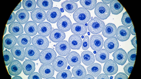
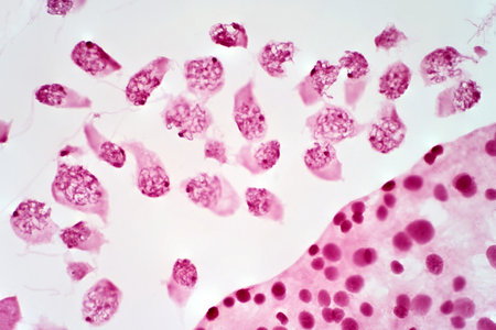
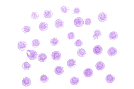
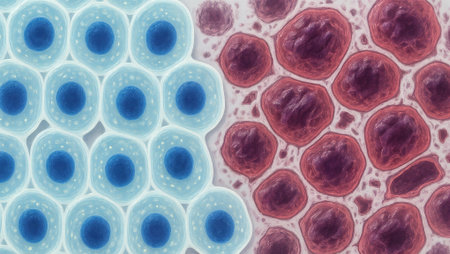
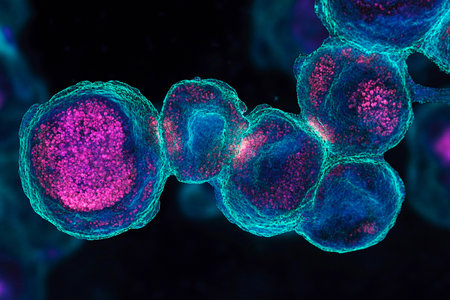
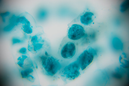
HeLa cervical cancer cells, stained with Coomassie blue, under microscope.
Коллекция по умолчанию
Коллекция по умолчанию
Создать новую

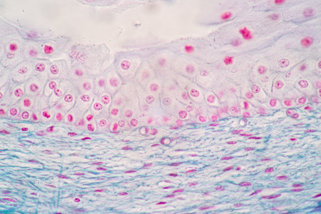
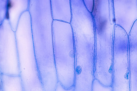
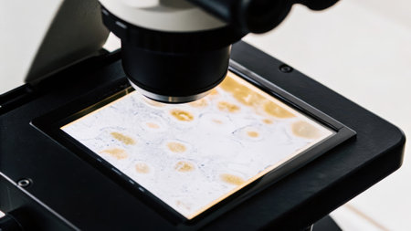
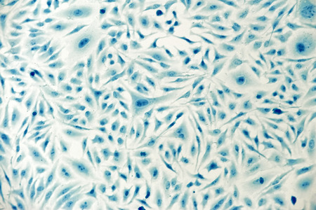
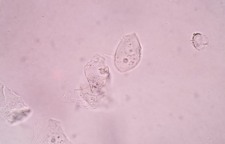
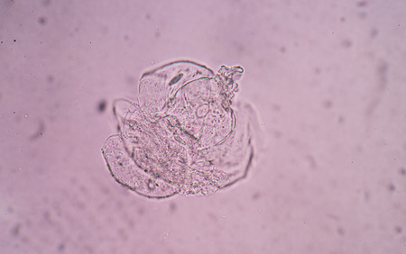
Characteristics of Squamous epithelial cell (Cell structure) of human under microscope view for education in laboratory.
Коллекция по умолчанию
Коллекция по умолчанию
Создать новую

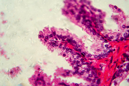
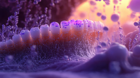
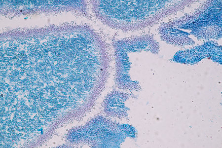
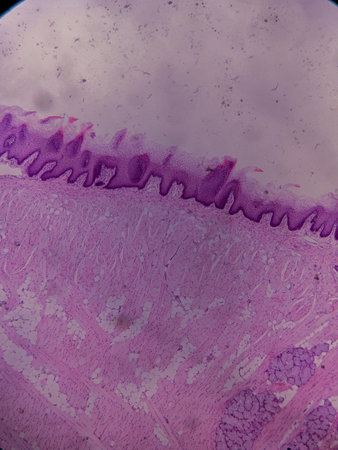
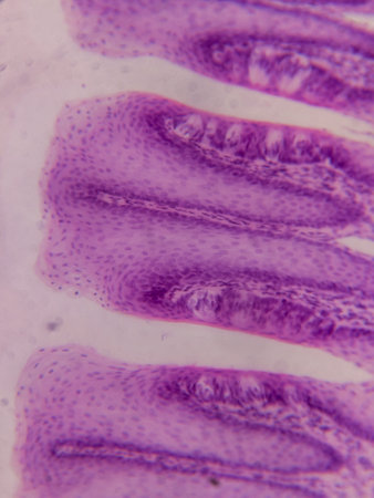
Tongue with taste buds Papilla across 100x
Коллекция по умолчанию
Коллекция по умолчанию
Создать новую

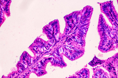
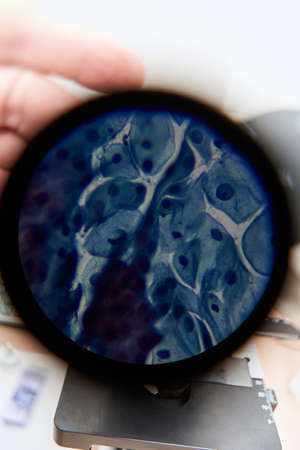
Stomach tissue under the microscope 100x
Коллекция по умолчанию
Коллекция по умолчанию
Создать новую
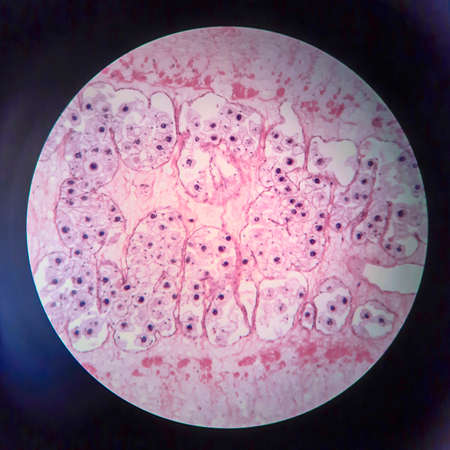
Pancreas cancer cells under microscope view for medical education.
Коллекция по умолчанию
Коллекция по умолчанию
Создать новую
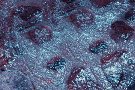
water flowing over boulders, rocks, and stones
Коллекция по умолчанию
Коллекция по умолчанию
Создать новую
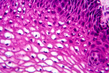
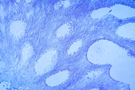
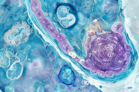
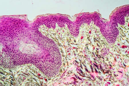
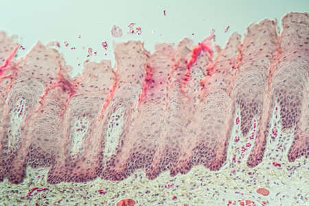
Atypical squamous epithelial hyperplasia, photomicrograph showing thickened stratified epithelium with nuclear atypia and basal layer crowding.
Коллекция по умолчанию
Коллекция по умолчанию
Создать новую
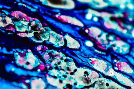
Blue and pink refill ink spilled onto the white washbasin and the ink mixed into abstract blobs and patterns.
Коллекция по умолчанию
Коллекция по умолчанию
Создать новую

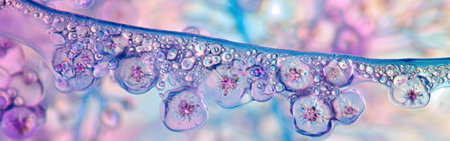
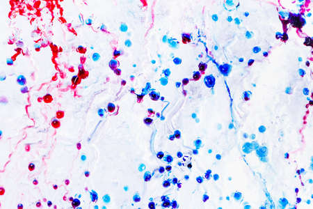
Cells in reproductive female cytology and histology education concept.
Коллекция по умолчанию
Коллекция по умолчанию
Создать новую
Showing Light micrograph of the Adrenal gland and Urinary bladder human under the microscope for education in the laboratory.
Коллекция по умолчанию
Коллекция по умолчанию
Создать новую
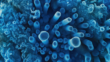
3d illustration of Neisseria gonorrhoeae bacteria cells
Коллекция по умолчанию
Коллекция по умолчанию
Создать новую
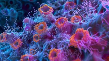
A highresolution image of tight junctions in epithelial cells displaying the various types and arrangements of proteins that make up these junctions
Коллекция по умолчанию
Коллекция по умолчанию
Создать новую
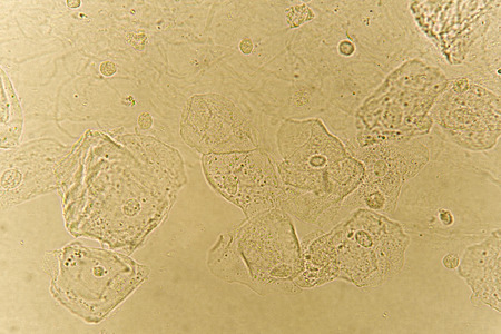
Epithelial cells with bacteria in patient urine
Коллекция по умолчанию
Коллекция по умолчанию
Создать новую

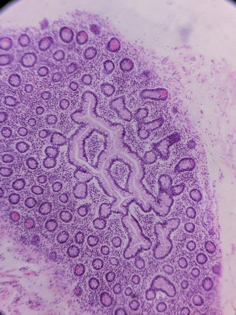
Histopathology of human under microscope view for education in laboratory.
Коллекция по умолчанию
Коллекция по умолчанию
Создать новую
Cross section of human cell under microscope view for education in laboratory.
Коллекция по умолчанию
Коллекция по умолчанию
Создать новую
Education anatomy and Histological sample of Human under the microscope.
Коллекция по умолчанию
Коллекция по умолчанию
Создать новую
Pancreas cancer cell under microscope view for medical education.
Коллекция по умолчанию
Коллекция по умолчанию
Создать новую
Pork tapeworm Taenia solium, section through the body, light micrograph
Коллекция по умолчанию
Коллекция по умолчанию
Создать новую
Microscopic view of human cells under microscope.
Коллекция по умолчанию
Коллекция по умолчанию
Создать новую

Calcium oxalate in urine find with microscope.
Коллекция по умолчанию
Коллекция по умолчанию
Создать новую
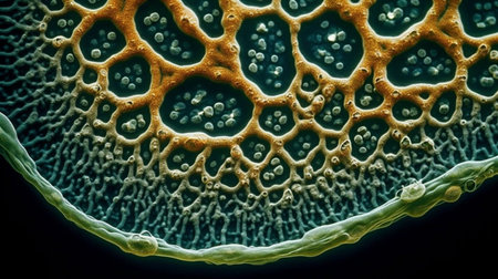
Close-up of plant stem cells under microscope, intricate patterns, high detail, with copy space
Коллекция по умолчанию
Коллекция по умолчанию
Создать новую
Close-up view of a textured surface featuring intricate patterns and vibrant blue tones, highlighting the beauty of natural materials and their unique formations
Коллекция по умолчанию
Коллекция по умолчанию
Создать новую
Education anatomy and Histological sample of Human under the microscope.
Коллекция по умолчанию
Коллекция по умолчанию
Создать новую
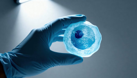
Gloved hand holds a translucent cell model under focused laboratory light with visible nucleus and organelle structures, suggesting scientific research and educational demonstration, with empty background space available for text
Коллекция по умолчанию
Коллекция по умолчанию
Создать новую
destructive mushroom in wood fabric 100x
Коллекция по умолчанию
Коллекция по умолчанию
Создать новую

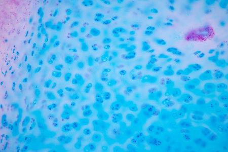
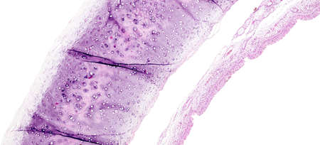
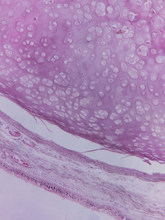
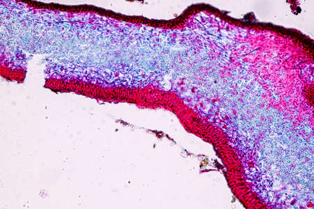
Human hyaline cartilage bone under microscope view for education pathology. Human tissue.
Коллекция по умолчанию
Коллекция по умолчанию
Создать новую

Tooth development from human under microscope view for education.
Коллекция по умолчанию
Коллекция по умолчанию
Создать новую
Macro shot of bubbles in water. Abstract background. Colorful background.
Коллекция по умолчанию
Коллекция по умолчанию
Создать новую
Hyaline cartilage, Elastic cartilage and Bone Human under the microscope in Lab.
Коллекция по умолчанию
Коллекция по умолчанию
Создать новую
Meiosis In Grasshopper Testis under the light microscope view.
Коллекция по умолчанию
Коллекция по умолчанию
Создать новую
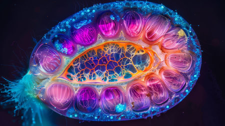
Vibrant cross-section of a developing seed under UV light, highlighting the embryo and endosperm in bright colors
Коллекция по умолчанию
Коллекция по умолчанию
Создать новую
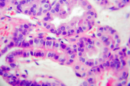
Photomicrograph showing histological features of benign prostatic hyperplasia. Enlarged prostate gland with nodular proliferation of glandular and stromal components. High-resolution histology image.
Коллекция по умолчанию
Коллекция по умолчанию
Создать новую
abstract of blur blue for background used
Коллекция по умолчанию
Коллекция по умолчанию
Создать новую
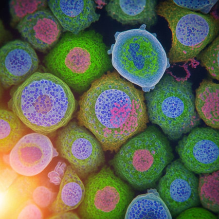
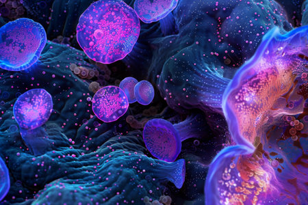
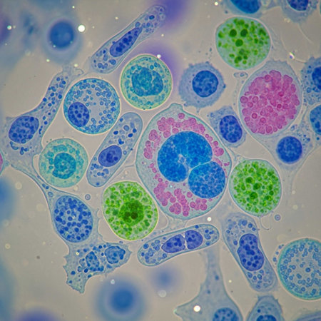
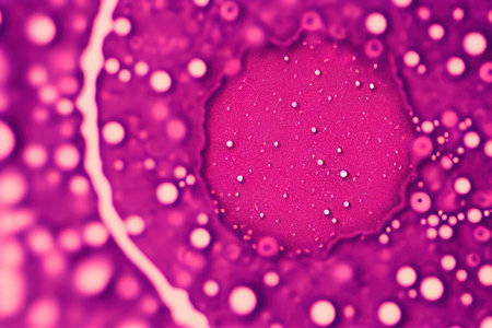
A colorful image of a cell with a purple and blue blob in the center. The image is abstract and has a mood of curiosity and wonder
Коллекция по умолчанию
Коллекция по умолчанию
Создать новую

Gloved hands hold a translucent cell culture sample with visible cellular structures and bubbles under focused clinical light, indicating laboratory research and microscopy, with available space for text on a neutral background
Коллекция по умолчанию
Коллекция по умолчанию
Создать новую
Exploring the microscopic world of cells and tissues, AI generated
Коллекция по умолчанию
Коллекция по умолчанию
Создать новую

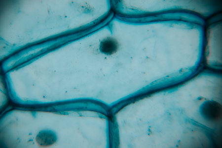
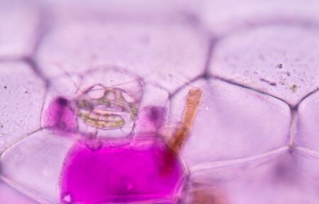
Close up Plant epidermis with stomata or Leaf Epidermis (Stomata) under microscope.
Коллекция по умолчанию
Коллекция по умолчанию
Создать новую
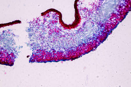
Study of protozoa and plant cells under the microscope for education.
Коллекция по умолчанию
Коллекция по умолчанию
Создать новую
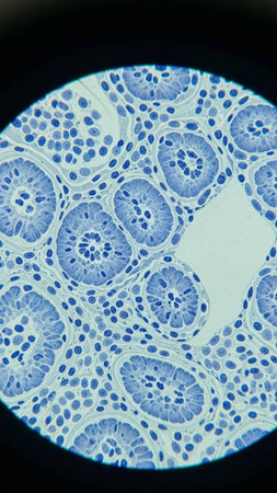
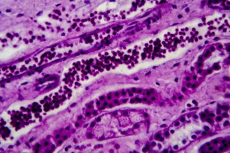
Columnar epithelium of human gall bladder under the microscope in Lab.
Коллекция по умолчанию
Коллекция по умолчанию
Создать новую

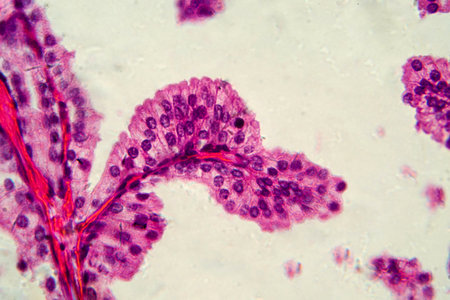
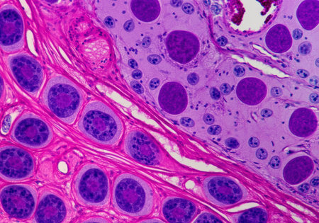
Learning anatomy and physiology of Pseudostratified columnar epithellum under the microscopic in laboratory. tissue,epithelium,columnar,biology,microscope,epithelial,microscopic,cells,cell,
Коллекция по умолчанию
Коллекция по умолчанию
Создать новую
A microscope eyepiece capturing the view of edited cells with a display showing realtime imaging where genetic changes can be seen highlighted in bright colors..
Коллекция по умолчанию
Коллекция по умолчанию
Создать новую

Proglottid (body unit) of tapeworm Taenia saginata, 3D illustration. A flatworm parasitizing animal and human intestine. Proglottid contains uterus with 12-30 primary lateral branches filled with eggs
Коллекция по умолчанию
Коллекция по умолчанию
Создать новую
abstract acrylic watercolor paint brush stroke texture isolated on white background for logo and banner. design, creative, and illustration.
Коллекция по умолчанию
Коллекция по умолчанию
Создать новую

A microscopic snapshot of a developing embryo with distinct patterns of histone modifications visualized as colorful spots crucial for regulating the expression of genes responsibl
Коллекция по умолчанию
Коллекция по умолчанию
Создать новую

Abstract background of a mix white blue paints, like a fantastic flowers
Коллекция по умолчанию
Коллекция по умолчанию
Создать новую
close up of a turquoise sofa texture for use as background
Коллекция по умолчанию
Коллекция по умолчанию
Создать новую
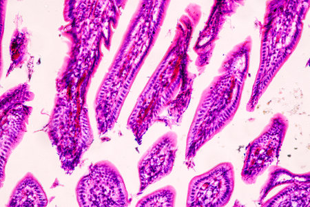
Tongue Tissue with taste buds across 200x
Коллекция по умолчанию
Коллекция по умолчанию
Создать новую
3d illustration of red blood cells with depth of field and bokeh
Коллекция по умолчанию
Коллекция по умолчанию
Создать новую
HeLa cervical cancer cells, stained with Coomassie blue, under microscope.
Коллекция по умолчанию
Коллекция по умолчанию
Создать новую
An abstract representation of dental microscopy features vibrant colors and intricate details, highlighting the beauty of biological structures under magnification and captivating viewers.
Коллекция по умолчанию
Коллекция по умолчанию
Создать новую
Beautiful image in microscope of microorganisms in the lab
Коллекция по умолчанию
Коллекция по умолчанию
Создать новую
Colorful microorganism cells close up. illustration in high details for medical education and science. ai generative.
Коллекция по умолчанию
Коллекция по умолчанию
Создать новую
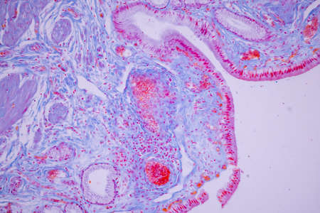
Histology of human tissue, show tracheitis as seen under the microscope
Коллекция по умолчанию
Коллекция по умолчанию
Создать новую

Ovarian cancer, light micrograph, photo under microscope. Photograph shows a fragment of a cancerous tumor in the female ovary. Selective focus
Коллекция по умолчанию
Коллекция по умолчанию
Создать новую
Tongue Tissue with taste buds across 200x
Коллекция по умолчанию
Коллекция по умолчанию
Создать новую
A close up of a blue and white cell structure, AI
Коллекция по умолчанию
Коллекция по умолчанию
Создать новую
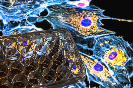
Achievements of modern biological science. A 24-well cell culture plate against an abstract biological background.
Коллекция по умолчанию
Коллекция по умолчанию
Создать новую
microscope lens, viewing Trypanosoma cruzi parasitic protozoan, causer of Chagas disease
Коллекция по умолчанию
Коллекция по умолчанию
Создать новую
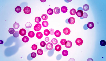
Blood cells in human body under microscope view for education in laboratory.
Коллекция по умолчанию
Коллекция по умолчанию
Создать новую
cell find with microscope.
Коллекция по умолчанию
Коллекция по умолчанию
Создать новую
Cell division under microscope view. Microscopic view of cell.
Коллекция по умолчанию
Коллекция по умолчанию
Создать новую
Human blood cells. 3D illustration showing the cells of the human blood cells
Коллекция по умолчанию
Коллекция по умолчанию
Создать новую
Characteristics of fungi living in wood as a group, are polyphyletic under the microscope for education.
Коллекция по умолчанию
Коллекция по умолчанию
Создать новую
Histopathology of human under microscope view for education in laboratory.
Коллекция по умолчанию
Коллекция по умолчанию
Создать новую
Photomicrograph showing histological features of benign prostatic hyperplasia. Enlarged prostate gland with nodular proliferation of glandular and stromal components. High-resolution histology image.
Коллекция по умолчанию
Коллекция по умолчанию
Создать новую
Diffuse proliferative glomerulonephritis, DPGN, light micrograph, photo under microscope. High magnification
Коллекция по умолчанию
Коллекция по умолчанию
Создать новую
Characteristics of Lichen, hyphae and Symbiotic algae under the microscope for education.
Коллекция по умолчанию
Коллекция по умолчанию
Создать новую
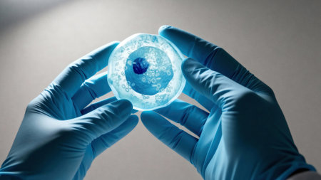
Gloved hands hold a translucent cell culture sample with visible cellular structures and bubbles under focused clinical light, indicating laboratory research and microscopy, with available space for text on a neutral background
Коллекция по умолчанию
Коллекция по умолчанию
Создать новую
Pink plant cells under microscope.400x
Коллекция по умолчанию
Коллекция по умолчанию
Создать новую
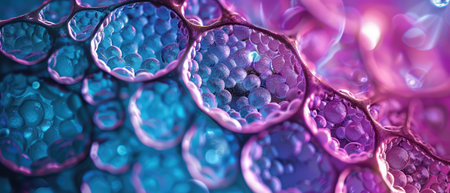
Colorful cells are showcased under a microscope, revealing their unique shapes and structures. The vibrant blues and purples illustrate complex biological interactions at a microscopic level.
Коллекция по умолчанию
Коллекция по умолчанию
Создать новую
Handmade modern abstract painting. Made with alcohol inks, by using watercolor techniques and rubbing alcohol. Useable as a background or texture.
Коллекция по умолчанию
Коллекция по умолчанию
Создать новую
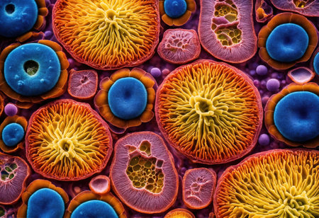
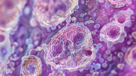
This detailed microscopic image showcases various cellular structures, highlighted in striking purple tones. The intricate patterns and textures reveal the complexity of biological tissues, making it a valuable resource for educational and scientific purposes
Коллекция по умолчанию
Коллекция по умолчанию
Создать новую

Epithelial cells with bacteria from the oral cavity 200x
Коллекция по умолчанию
Коллекция по умолчанию
Создать новую
Microscope with metal lens at laboratory. Medical equipment.
Коллекция по умолчанию
Коллекция по умолчанию
Создать новую
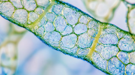
Explore the intricacies of plant cells through this detailed microscopic image, showcasing their unique structure and transparency. Perfect for scientific studies.
Коллекция по умолчанию
Коллекция по умолчанию
Создать новую
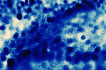
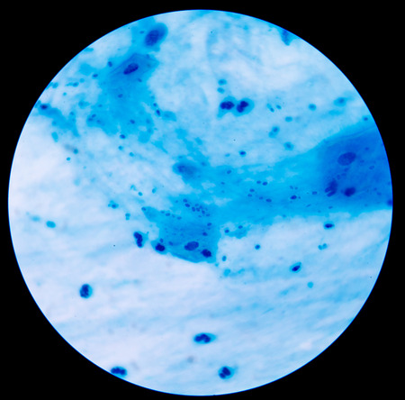
Microscopic image of blue-stained cells with different shapes and sizes on a light background, displaying cellular structures and details in laboratory conditions
Коллекция по умолчанию
Коллекция по умолчанию
Создать новую
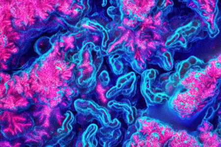
Thermal photography of an abstract, colorful pattern with heat waves in the style of Matisse. The color palette features neon pink and blue, with vivid colors. Captured using a macro lens, the image has an x-ray effect and a dynamic composition. Created using a redshift renderer, the result is hyper-realistic, with detailed textures and a cinematic, high-resolution appearance. --ar 3:2 --v 6.1 Job ID: 06163584-6a82-45d4-b422-f688ac3881ef
Коллекция по умолчанию
Коллекция по умолчанию
Создать новую

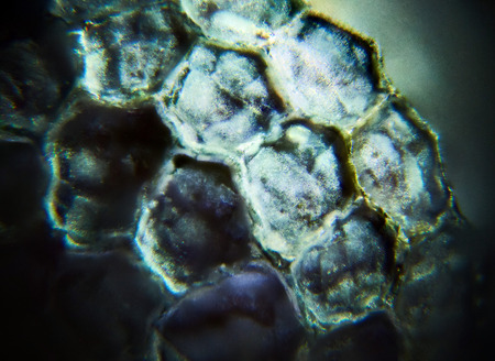
Apple pollen from a blossom in spring under the microscope
Коллекция по умолчанию
Коллекция по умолчанию
Создать новую
Papillary thyroid carcinoma, light micrograph, photo under microscope. The most common type of thyroid cancer
Коллекция по умолчанию
Коллекция по умолчанию
Создать новую
Histopathology of human under microscope view for education in laboratory.
Коллекция по умолчанию
Коллекция по умолчанию
Создать новую
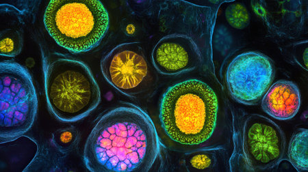
Vivid microscopic imagery showcasing intricate cellular structures illuminated in various colors, revealing the beauty of life at a microscopic level.
Коллекция по умолчанию
Коллекция по умолчанию
Создать новую
Spread-out acrylic paint. Abstract background, made in the technique of fluid art. Demonstrating the colors of 2023 - Viva Magenta
Коллекция по умолчанию
Коллекция по умолчанию
Создать новую
squamous cell
Коллекция по умолчанию
Коллекция по умолчанию
Создать новую
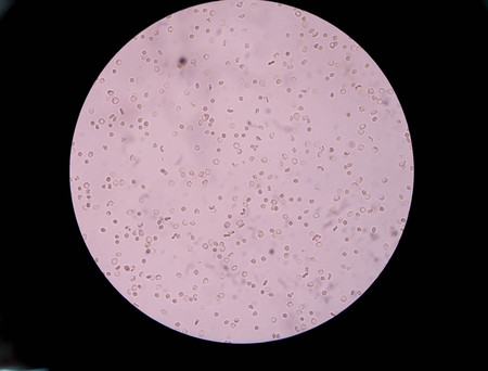
Red blood cell in urine under microscopic
Коллекция по умолчанию
Коллекция по умолчанию
Создать новую
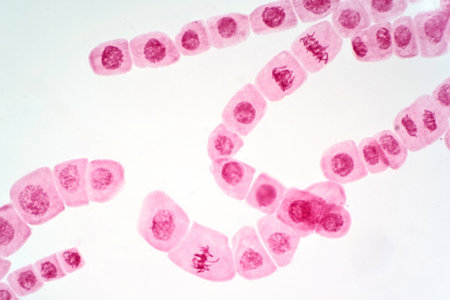
Mitosis cell of root tip of onion under the light microscope view.
Коллекция по умолчанию
Коллекция по умолчанию
Создать новую

Abstract macro image of particles looking like bacteria, macro shot, microbiology theme
Коллекция по умолчанию
Коллекция по умолчанию
Создать новую
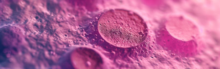
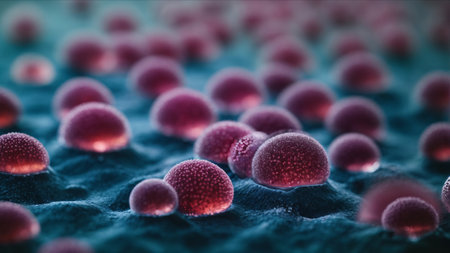
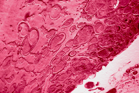
Abstract pink cancer cell organism background 3d render digital illustration
Коллекция по умолчанию
Коллекция по умолчанию
Создать новую
Microscopic view of tissue sections showing purple-stained cells, possibly from a biological sample
Коллекция по умолчанию
Коллекция по умолчанию
Создать новую
Close up of water drops on a pink background. Abstract background.
Коллекция по умолчанию
Коллекция по умолчанию
Создать новую
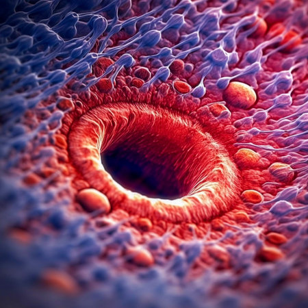
3D illustration of a microscopic view of a human cell under microscope.
Коллекция по умолчанию
Коллекция по умолчанию
Создать новую
Chaos ink texture background, ink in water pattern frost. Crystal winter design
Коллекция по умолчанию
Коллекция по умолчанию
Создать новую
Viscous thick oil texture with industrial dark background showcasing vibrant patterns
Коллекция по умолчанию
Коллекция по умолчанию
Создать новую
Characteristics of Lichen, hyphae and Symbiotic algae under the microscope for education.
Коллекция по умолчанию
Коллекция по умолчанию
Создать новую
Microscopic seeds opium poppy Papaver somniferum. Wet shoots by microscope. Narcotic, drug opiates and food plant. Super macro close-up
Коллекция по умолчанию
Коллекция по умолчанию
Создать новую
Extremely closeup of human cell, view through a microscope, surreal illustration, AI Generated
Коллекция по умолчанию
Коллекция по умолчанию
Создать новую
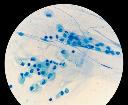
Branching budding yeast cells with pseudohyphae in sputum gram stain fine with microscope.
Коллекция по умолчанию
Коллекция по умолчанию
Создать новую
black textured surface covered with countless tiny white specks
Коллекция по умолчанию
Коллекция по умолчанию
Создать новую
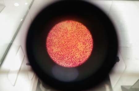
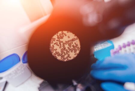
Smear of Acid-Fast bacilli AFB stained from sputum specimen, under 100X light microscope.
Коллекция по умолчанию
Коллекция по умолчанию
Создать новую

Characteristics of Lichen, hyphae and Symbiotic algae under the microscope for education.
Коллекция по умолчанию
Коллекция по умолчанию
Создать новую
A closeup of glandular epithelium highlighting its specialized cells for secretion and absorption
Коллекция по умолчанию
Коллекция по умолчанию
Создать новую
Legion-Media
Создайте свои проекты на основе качественных стоковых фотографий и видео.


Copyright © Legion-Media.